Views: 0 Author: Site Editor Publish Time: 2025-07-24 Origin: Site


You might remember when you first saw an x-ray. You may have been surprised by the bright white areas called "shadows." These white spots show up because dense things like bone block more x-rays. This means fewer rays reach the film or detector. Long ago, doctors thought these shadows were just odd shapes. They did not think they were important tools. Now you know these shadows show important details. Many people think shadows on x-rays always mean sickness. But normal body parts or old injuries can make them too. Knowing this helps you understand x-ray images better.
Dense materials like bone block more X-rays, making those areas appear white on the image.
Soft tissues let more X-rays pass through, so they show up as gray shades on an X-ray.
Air spaces, such as lungs, do not block X-rays and appear black on the film.
The white 'shadows' on an X-ray are bright spots where fewer X-rays reach the detector.
Understanding these shadows helps you read X-rays better and feel more confident about your health.
When you get a radiograph, a special machine sends x-ray energy through your body. Different tissues react in their own ways. Bones, muscles, and fat all interact with x-rays. Collagen in bone can change or break down if the dose is high. Soft tissues, like muscle, may form tiny bubbles if water splits apart. These changes can make a radiograph look less clear, especially in high-resolution medical imaging.
Tip: Remember, radiographs use much lower doses than those that cause damage. Doctors keep you safe by using the lowest dose needed for clear images.
You see different shades on a radiograph because tissues absorb x-rays in different ways. Dense tissues, like bone, have more atoms with higher atomic numbers. This means they block more x-rays. Soft tissues, like muscle and fat, let more x-rays pass through. This gives a range of shades from white to black on your radiograph.
Here's a table showing how bone and soft tissue are different:
Aspect | Bone | Soft Tissue | Effect on Radiograph |
|---|---|---|---|
Atomic Number | High (calcium, phosphorus) | Low (hydrogen, carbon, oxygen) | Bone appears white |
Density | High | Low | Soft tissue appears gray |
X-ray Interaction | Photoelectric effect dominates | Compton scatter dominates | Stronger absorption in bone |
Imaging Outcome | White/opaque | Gray or dark | Clear contrast in medical imaging |
Radiographs work because of these differences. The photoelectric effect depends on atomic number and is important in medical imaging. Bones absorb more energy, so they show up as bright white. Soft tissues absorb less, so they look gray. Air spaces, like in your lungs, look darkest. This contrast helps doctors find problems quickly and safely during medical imaging.
When you look at a radiograph, you see bright white areas where dense materials block the x-ray beam. Bone stands out because it contains a lot of minerals, especially calcium carbonate. This mineral content gives bone a higher atomic number than soft tissue. It means bone absorbs more x-rays through a process called photoelectric absorption. Soft tissue, like muscle or fat, has fewer minerals and a lower atomic number. It lets more x-rays pass through.
You can think of bone as a strong wall. It stops most of the x-rays from reaching the film or detector. Soft tissue acts more like a thin curtain. It lets many x-rays go by. The difference in how much x-rays get blocked creates the patterns you see on a radiograph.
Note: The density of a material changes how much it blocks x-rays. Scientists have tested this using different materials. For example, a thick iron plate blocks almost all x-rays, while a thin sheet of paper lets most pass through. The denser the material, the more it shows up as a white area on your radiograph.
The white areas you see on a radiograph are called "shadows on x-ray." These shadows appear because fewer x-rays reach the detector in those spots. When bone or another dense object blocks the x-rays, the film does not get exposed as much. Less exposure means a brighter, whiter spot on the image.
Radiologists use the term "shadow" to describe these white outlines. The shadow is not a dark area like you see when you block sunlight. Instead, it is a bright area where x-rays could not pass through. The size and sharpness of the shadow depend on how far the object is from the x-ray source and the detector. Radiologists measure the shadow's size using a formula that compares the distance from the x-ray source to the image and the distance from the source to the object. They also look at how clear the edges are, which can change if the x-ray beam spreads out or if the object is not close to the detector.
Tip: The brightness of the shadow on a radiograph depends on how many x-rays hit the film. If you increase the x-ray exposure, the film gets darker. If you block more x-rays, the film stays lighter. Hospitals use special machines to control the exposure and keep the images clear.
Clinical studies show that radiologists can read these shadows on x-ray with high accuracy. New computer tools help them spot important changes even faster. These tools do not replace the doctor but make it easier to find problems in the white shadows you see on your radiograph.
Key Points to Remember:
Bone blocks more x-rays than soft tissue because it is denser and has more minerals.
The white "shadows on x-ray" show where x-rays could not reach the detector.
Radiologists use the size and sharpness of these shadows to help diagnose problems.
The brightness of the shadow depends on how many x-rays get blocked.
When you look at a radiograph, you see three main colors: white, gray, and black. Each color tells you something about what is inside the body. White areas show up where dense materials block x-rays. Bone is the most common example, but metal and contrast agents also appear white. These materials absorb x-rays strongly, so almost no rays reach the detector in those spots.
Gray areas on a radiograph represent soft tissue. Muscle, fat, and organs like the liver or heart fall into this group. These tissues let some x-rays pass through, but not as many as air. That is why they look gray instead of black or white.
Black areas show where air fills a space. Your lungs are a good example. Air does not block x-rays, so the detector gets the most exposure in these spots. This makes them appear darkest on the radiograph.
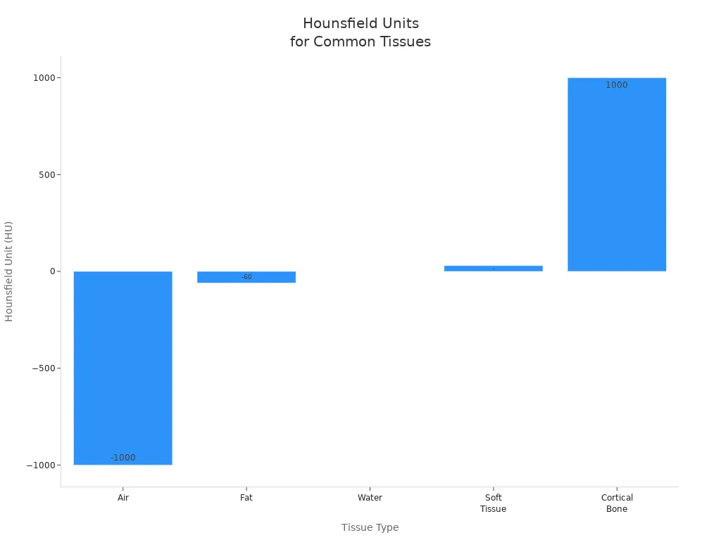
Here is a table that shows how different tissues look on a radiograph and their typical grayscale values:
Tissue Type | Typical Hounsfield Unit (HU) | Appearance on Radiograph |
|---|---|---|
Air | -1000 | Black |
Fat | -60 | Dark gray |
Soft Tissue | 20 to 40 | Gray |
Water | 0 | Mid-gray |
Cortical Bone | +1000 or more | White |
Metal/Contrast | >+1000 | Bright white |
You can also see these differences in the chart below:

You can use these colors to figure out what you see on a radiograph. White means bone, metal, or a contrast agent. Gray means soft tissue, like your heart or muscles. Black means air, such as in your lungs or intestines.
If you see a bright white spot, it could be a bone or a piece of metal.
A gray area might be your liver, kidney, or muscle.
A black area usually means air, like in your lungs.
Radiologists use these shades to find problems. They look for changes in the normal pattern of white, gray, and black. For example, a white area in the lung could mean pneumonia or a tumor. A black area where bone should be might mean a fracture.
Tip: In medical imaging, radiologists rely on their ability to spot small changes in these shades. They use their training to read radiographs and find signs of disease or injury. Digital radiographs show up to 256 shades of gray, so even tiny differences can matter.
You now know how to read the basic colors on a radiograph. This skill helps you understand what doctors see during medical imaging. It also helps you ask better questions about your own health.
You see shadows every day when sunlight or a lamp shines on objects. These shadows form because something blocks the light. Your hand, a tree, or a wall can stop visible light, making a dark shape on the ground or wall. Visible light has longer wavelengths and lower energy than X-rays. It cannot pass through most solid things.
Let's look at how visible light and X-rays compare:
Property | Visible Light | X-rays |
|---|---|---|
Wavelength | 400 to 700 nanometers | 0.03 to 3 nanometers |
Energy | Lower energy photons | Much higher energy photons |
Shadow Formation | Shadows formed by blocking or absorption of light by opaque objects | Shadows formed by differential absorption based on density (e.g., bones absorb more X-rays than skin) |
Penetration Ability | Limited to surface or opaque objects | Can penetrate materials like human tissue and bones |
X-rays act very differently. They have much shorter wavelengths and higher energy. This means they can travel through your skin and soft tissue. Only dense things, like bone or metal, can stop them. That is why you do not see regular shadows on x-ray images. Instead, you see patterns based on how much each part of your body absorbs the X-rays.
Think about holding your hand in front of a flashlight. You see a dark shadow because your hand blocks the light. Now, imagine X-rays instead of light. X-rays pass through most of your hand, but your bones stop more of them. On an X-ray image, the places where bones block X-rays show up as white areas. These are the shadows on x-ray.
You can picture it like this:
Light shadows: Dark shapes where light cannot pass.
X-ray shadows: White shapes where X-rays cannot pass.
The difference comes from how each type of energy interacts with matter. Visible light cannot go through your hand, so you get a dark shadow. X-rays can go through soft tissue but not bone, so you get a white shadow where the bone is. This is why shadows on x-ray look white, not black.
Tip: Next time you see an X-ray, remember that the white areas are not empty spaces. They show where something dense, like bone, stopped the X-rays from reaching the detector.
Dense materials, like bone, block X-rays and create the white 'shadows' you see on the film. Knowing this helps you understand your own or your pet's X-rays. When you see these images, you feel more confident and less anxious because you can spot what is normal and what is not.
You get clearer answers when doctors or veterinarians use simple words and pictures.
Seeing your X-ray helps you feel involved and in control.
Good explanations make you less worried and more prepared for decisions.
Remember: Learning how to read X-ray images gives you power to ask questions and take part in your care.
Bones block most X-rays. The detector receives fewer rays in those spots. It records these areas as white. You see bone as a bright shape because it stops the X-rays from passing through.
You can see soft tissues, but they look gray. They let some X-rays pass through. The detector picks up more rays than with bone, but fewer than with air. This creates a gray shade.
Air in your lungs does not block X-rays. The detector gets almost all the rays in those areas. It records them as black. You see your lungs as dark spaces on the image.
Metal blocks nearly all X-rays. It appears as a very bright white spot. Doctors can spot metal easily because it stands out from bone and soft tissue.
Tip: If you have metal in your body, tell your doctor before an X-ray. It helps them read your image correctly.