Views: 0 Author: Site Editor Publish Time: 2025-08-26 Origin: Site


You help get clear images when you use the right positioning in veterinary dental radiography. This keeps patients safe and helps get correct results. Here is why it matters:
Aspect | Importance |
|---|---|
Diagnostic Accuracy | Getting the same images each time helps doctors understand dental problems. |
Patient Safety | Using general anesthesia during radiographs keeps both patients and staff safe. |
When you use advanced veterinary equipment from SHINOVA, you work faster and get better results in radiography.
Good positioning helps get clear dental pictures. Make sure the patient and x-ray beam line up right. This helps the images show what is needed.
Safety is very important when taking x-rays. Wear protective gear and follow safety rules. This keeps patients and staff safe.
Check the patient well before taking x-rays. This helps you pick the best position. It also means you will not need to retake pictures.
Pick the right sensor size for each animal. The correct sensor gives better pictures. It also lowers the amount of radiation.
Try different ways to position animals. Change your method for each animal's size and age. This helps you get the best results.
You need clear and correct images every time. Good positioning helps you see the teeth and jaw well. This makes it easier to find dental problems early. You must line up the patient and the x-ray beam the right way. This step helps you follow the rules for dental radiography. Using the right positioning keeps the patient safe and gives the best results.
How you position the patient is very important for clear images.
The x-ray beam must be lined up right for good pictures.
How long you take the x-ray changes how clear the image is.
Doing things the right way helps you get better results.
SHINOVA gives you advanced veterinary equipment. These tools help you position things right and get good results.
You need to keep the patient and your team safe. Always follow safety rules during radiography. Wear gear like lead aprons and thyroid shields. Use badges to check how much radiation you get. Ask your team to leave the room when you scan. Stay far from the x-ray source.
Safety Protocols | Description |
|---|---|
Protective Equipment | Use protective gear for the mouth, skin, eyes, ears, and breathing. |
Irrigation | Rinse the mouth with 0.12% chlorhexidine to lower germs in the air. |
Radiation Protection | Wear lead aprons, thyroid shields, and use radiation badges. |
Room Occupancy | Do not stand in the x-ray beam and keep a safe distance. |
Ergonomics | Sit and position yourself right to avoid getting hurt. |
You can count on SHINOVA for safe and high-quality equipment.
You need to think about many things to get the best images. Your skill as an operator is very important. The animal's skull size and shape can change how you position them. The settings on your machine also matter. Practice often to get better at this. SHINOVA's new tools help you do your best in dental radiography positioning.

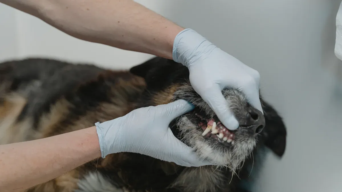
You start by checking the patient before any dental radiography. It helps you get the best results. You look at the animal's health and mouth. You choose the right position for the head and jaw. You use aids like sand bags or troughs to keep the patient steady. You place the film so it shows the whole tooth and root. You pick the film size based on the animal's size. You set the beam head so it points at the area you want to see.
Tip: Careful assessment lowers the chance of retakes and saves time.
Steps for Pre-Radiograph Assessment:
Position the patient using supports.
Place the film to capture the full tooth and root.
Adjust the beam head for clear images.
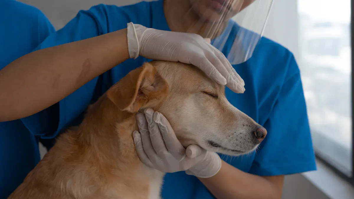
You keep the patient calm and still during the procedure. You use sedation to lower stress and stop movement. You pick the right drugs for each animal. You combine drugs for better results. You use gentle restraint to keep the patient safe.
Sedation Protocol | Effects on Sedation and Analgesia | Combination Benefits |
|---|---|---|
Dexmedetomidine + Fentanyl | Sedation and moderate pain relief | Works well with opioids and benzodiazepines |
Xylazine + Fentanyl | Good pain relief and sedation | Helps relax muscles for radiography |
Note: You always watch the patient during sedation. You adjust the dose as needed.
You help the patient feel safe and relaxed. You talk to clients about anesthesia to lower their fears. You teach them about the procedure. You use dental health report cards to show the pet's oral health. It helps clients trust the process and follow your advice.
You answer questions about anesthesia for older pets.
You explain each step to lower anxiety.
You share dental health report cards for better understanding.
You make patient preparation smooth and stress-free. It leads to better images and safer procedures in veterinary dental radiography.
Setting up your equipment the right way helps you get clear images. SHINOVA gives you advanced tools and support. This makes your work easier and more dependable.
You must pick the right sensor for each patient. Digital radiography gives sharp pictures and shows small details. You can change brightness, contrast, and zoom to see better. You get images fast, so you can help patients sooner. Less radiation keeps everyone safer.
Film Type | Radiation Exposure | Resolution Impact | Recommendation |
|---|---|---|---|
E Speed | Less than D Speed | Slight decrease | Good for experienced practitioners |
F Speed | Less than E Speed | Slight decrease | Good for experienced practitioners |
D Speed | Higher | Higher | Good for beginners |
Pick a sensor size that fits the animal's mouth. Using size 2 or size 4 sensors helps you see the whole tooth and root. Always check where the sensor is before taking a picture.
Less radiation for safety
Fast images for quick answers
Easy changes for clearer pictures
You need to put the x-ray generator in the best place. For maxillary teeth, lay the patient on their chest. For mandibular teeth, lay them on their back. Put the sensor inside the mouth under the tooth you want to see. Point the tube head straight at the angle between the tooth and sensor.
Recommendation | Description |
|---|---|
Range of Motion | Use a generator that moves for different angles |
Positioning for Size 2 Plates | Keep the generator close to the patient |
Positioning for Larger Plates | Move the generator farther to cover the full plate |
Tip: Keep the x-ray head about 1-2 inches from the fur. This helps stop blurry pictures.
Set the exposure right for each patient. Use the lowest dose that still gives a clear image. Change settings for the animal's size and the area you need to see. SHINOVA's tools let you change settings fast and safely.
New tools like intraoral CMOS sensors give better images. They also make positioning easier. You can trust these tools to help you do your best work.

You need to follow clear steps for maxilla positioning. This helps you get sharp images for the upper jaw. Use this dental positioning guide for both dogs and cats.
Place the patient in sternal recumbency. Prop a towel under the chin. This keeps the maxilla parallel to the table.
Position the digital sensor flat against the teeth. Keep it parallel to the table.
For the first and second maxillary molars, place the sensor at the back edge of the second molar. Set the x-ray tube at a 60-degree angle. Aim high and toward the back.
For the fourth premolar, move the sensor forward. Adjust the tube to 50 degrees. Aim straight at the tooth.
Move the sensor forward again for the premolars. Set the tube at 45 degrees. Aim high.
For the maxillary canine, use your fingers to make an L shape for guidance. Set the tube at 70 degrees. Aim at the tip of the canine tooth.
To image the maxillary incisors, move the sensor to the front. Angle the tube at 45 degrees. Center it in front of the incisors.
Tip: Use a size 2 sensor for cats and small dogs. Use a size 4 sensor for large dogs. This helps you capture the full mouth series.
Mandible positioning helps you see the lower jaw. You need to follow these steps for clear radiographs.
Place the patient in dorsal recumbency for mandibular incisors and canines. For premolars and molars, use lateral recumbency.
Position the sensor inside the mouth under the tooth you want to image.
For mandibular molars and premolars, keep the sensor parallel to the teeth. Place it as far back as possible.
For the mandibular canines and incisors, move the sensor forward. Angle it to fit the curve of the jaw.
Adjust the x-ray tube so it points straight down for parallel technique. For bisecting angle, tilt the tube to match the angle between the tooth and sensor.
Quick reference: Use sandbags or foam pads to keep the head steady during the procedure.
The parallel technique works best for mandibular premolars and molars in dogs. It gives you accurate images without distortion.
Steps for the Parallel Technique:
Place the patient in lateral recumbency.
Insert the sensor inside the mouth. Keep it parallel to the teeth.
Position the x-ray tube head so the beam is perpendicular to both the sensor and the teeth.
Take the radiograph. Check the image for clarity.
Tooth Type | Best Technique | Sensor Size |
|---|---|---|
Mandibular Molars | Parallel | Size 2 or 4 |
Mandibular Premolars | Parallel | Size 2 or 4 |
Maxillary Teeth | Bisecting Angle | Size 2 or 4 |
Note: The parallel technique is a routine procedure for full mouth radiographs in large dogs.
You use the bisecting angle technique for maxillary teeth and some mandibular teeth in cats and small dogs. This helps you avoid image distortion.
Steps for the Bisecting Angle Technique:
Place the patient in lateral recumbency. Use a 3 cc syringe cover as a mouth gag.
Insert a size 4 intraoral film. Keep one side against the palatal margin.
Bend the other edge of the film. Tuck it behind the lower first molar to hold it in place.
Adjust the film to create a small space between it and the palate. Try to keep it near parallel to the root of the tooth.
Position the x-ray tube head. The central beam should be at a right angle to the upper part of the film and parallel to the muzzle.
For canine teeth, move the film so the back edge sits in front of the fourth premolar. Tip the front edge to line up with the canine tooth. Make sure the tube head matches this angle.
Tip: The bisecting angle technique is key for full mouth series in cats and small dogs. It helps you get clear dental x-rays for all teeth.
You can use this veterinary dental radiographic positioning guide as a quick reference for every dental procedure. It helps you get the best results for full mouth radiographs. SHINOVA equipment supports you in every step of veterinary dental radiography. You can trust this guide for both routine procedure and advanced cases.
You work with small animals a lot in dental radiography. These animals can move around easily. You need to be careful with them.
Use manual restraint for short x-ray exams if the animal is calm.
Use chemical restraint for longer or harder procedures. Sedatives or inhalant anesthetics help keep the animal still.
Pick the right amount of medicine for each kind of animal.
Use small troughs or tape to hold their legs. Heavy sandbags are not good for small animals.
Check how the animal is lying before each picture.
Watch for movement or teeth that overlap in the x-ray. If you see this, move the animal or sensor and try again.
Tip: Calm and gentle handling helps you get clear pictures and keeps the animal safe.
Large dogs need strong support for dental x-rays.
Use foam pads or sandbags to keep the head and body still.
Put the sensor deep in the mouth to see big teeth.
Ask someone to help hold the dog if you need it.
Check the jaw and sensor to make sure they line up before you take the x-ray.
Look for teeth that overlap in the picture. Move the sensor or change the angle if you see this.
Common Issue | Solution |
|---|---|
Movement | Use more restraint or sedation |
Overlapping teeth | Change the sensor's angle or spot |
Young and old animals need extra care when you position them.
For older animals, watch their organs closely.
Use anesthesia that helps the heart and lungs.
Keep the animal warm so their body temperature does not drop.
Add soft padding to protect thin or weak bodies from sores.
For young animals, use gentle restraint and soft supports.
Always check that the animal is comfortable and safe before you start.
Note: Careful changes for age help you avoid injury and get better results in dental radiography.
You may see images that look stretched or squished. This happens when the x-ray beam does not hit the teeth at the right angle. If you miss the tips of the teeth, you may need to change the vertical angle. Too much angle can make teeth look short. Too little angle can make them look long. Always check the sensor placement. Make sure it covers the area you want to see. Handle the sensor gently. Bends or folds can cause strange marks on the image.
Error | What You See | How to Fix It |
|---|---|---|
Foreshortening | Teeth look short | Lower the vertical angle |
Elongation | Teeth look long | Raise the vertical angle |
Overlapping Contacts | Teeth overlap each other | Adjust the horizontal angle |
Cut-off Apices | Tooth tips missing | Move the sensor or change the angle |
Tip: Place the sensor flat and keep the x-ray beam perpendicular to the bisecting angle. This helps you avoid distortion.
Animals may move during the x-ray. Movement can blur the image. You can use gentle restraint or sedation. Check the animal's comfort before you start. Use foam pads or towels to keep the head steady. Watch for signs of stress. If you see movement, pause and adjust your setup. Practice helps you get better at keeping animals still.
Use soft supports for comfort.
Sedate if needed for longer exams.
Check the animal's position before each shot.
Repeat the image if you see blur.
Cone cuts show up as blank spots on the image. This means the x-ray beam missed part of the sensor. Overlapping happens when teeth cover each other in the picture. You can fix these problems by checking your tube and sensor placement.
Problem | Solution |
|---|---|
Cone Cut | Center the x-ray beam over the sensor. Move the tube toward the blank area. |
Overlapping | Adjust the horizontal angle. Aim the beam between the teeth. |
Film Cut-off | Shift the sensor toward the area that is missing. |
Note: For best results, always check your positioning before you take the x-ray. In veterinary dental radiography, small changes can make a big difference.
You can get clear pictures in veterinary dental radiography by doing a few important things.
Put the patient in the correct spot.
Turn the head so parts do not cover each other.
Use a special prop to keep the mouth open.
Tilt the x-ray tube to see better.
Mark the area you want to check for better accuracy.
Doing these steps the same way each time gives you sharp pictures and helps stop mistakes. Many veterinary classes let you practice and listen to lessons to get better. SHINOVA gives you tools and help so you can do your best work every day.
You need to put the sensor in the right spot. Make sure the x-ray beam lines up with the sensor. This helps you get clear pictures. It also keeps the patient safe.
You can use gentle restraint or sedation. Soft things like foam pads help keep the animal still. Always check if the animal feels comfortable before you start.
Blurry or weird pictures happen when the x-ray beam does not hit the sensor at the right angle. You should check where the sensor is and fix the beam before you take the x-ray.
Digital sensors and advanced x-ray generators help you get sharp pictures. SHINOVA has tools that make positioning easier and help you get better results in veterinary dentistry for the veterinary technician.
No. You need to change your technique for each animal's size and age. Small animals, big dogs, and older pets need different supports and angles for the best results.